42 human eye with labels
labeled structure of human eye Eye Model Labeled - Bing Images | Human Anatomy And Physiology, Eye . eyeball ojo. 1000+ Images About Medical Information Eyes On Pinterest | Eye Anatomy . eye anatomy eyes human. Human Brain Internal Structure mandevillehigh.stpsb.org. How to draw the Human Eye - Labeled Science Diagrams - YouTube Download a free printable outline of this video and draw along with us: you for watching. Please subsc...
eye labelled simple - anatomyschool.z21.web.core.windows.net eye labelled simple. MP Board Class 10 Science 2017 Previous Year Question Paper And. 16 Images about MP Board Class 10 Science 2017 Previous Year Question Paper And : Blank Eye Diagram - Cliparts.co, Parts of the Eye Diagram for 4th graders | Lesson 2 Grade 3 - Grade 4 and also Human Eye Eye Diagram Not Labeled - Aflam-Neeeak.

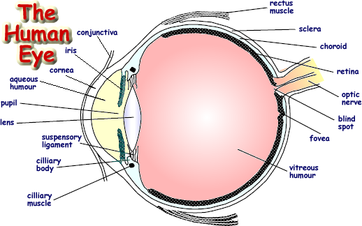
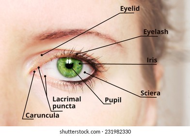
Human eye with labels
The Eyes (Human Anatomy): Diagram, Optic Nerve, Iris, Cornea ... - WebMD The front part (what you see in the mirror) includes: Iris: the colored part. Cornea: a clear dome over the iris. Pupil: the black circular opening in the iris that lets light in. Sclera: the ... Structure and Functions of Human Eye with labelled Diagram - BYJUS The human eye is a roughly spherical organ, responsible for perceiving visual stimuli. It is enclosed within the eye sockets in the skull and is anchored down by muscles within the sockets. Anatomically, the eye comprises two components fused into one; hence, it does not possess a perfect spherical shape. Human eye model labeled Flashcards | Quizlet all of the open space. Lacrimal Gland. Fovea centralis. the black dot in the middle. Medial Commissure. Lacrimal Ducts. Macula Lutea. The pink dot (not the black in the middle)
Human eye with labels. Labeled Eye Diagram - Pinterest Labeled Eye Diagram. Samer kursa. 16 followers. Eye Anatomy Diagram. Human Eye Diagram. Diagram Of The Eye. Brain Anatomy. Anatomy And Physiology. Human Anatomy. Anatomy Organs ... Human Eye. This vibrant 20" x 26" (51 x 66 cm) exam-room anatomy poster shows cross section of The Eye. It also provides lateral and superior view of the eye and ... Human Eye Diagram, How The Eye Work -15 Amazing Facts of Eye Fun Facts About Human Eye For Kids FACT 1 Iris scanning is more secure than fingerprints because our iris has 256 unique characteristics and the fingerprint has just 40. FACT 2 Newborn babies don't produce tears. They only make crying sounds, but no tears come out of their crying eyes. Labeled Eye Diagram | Science Trends What you want to interpret as a major part of the human eye is somewhat up to the individual, but in general there are seven parts of the human eye: the cornea, the pupil, the iris, the lens, the vitreous humor, the retina, and the sclera. Let's take a closer look at each of these components individually. The Cornea diagram of eye with labels Horseshoe Crab Anatomy. 16 Pics about Horseshoe Crab Anatomy : Label the Eye, Eye With Labels Clip Art at Clker.com - vector clip art online, royalty and also Muscles of the Human Eyeball | ClipArt ETC. Horseshoe Crab Anatomy dnr.maryland.gov crab horseshoe anatomy eyes diagram labeled gills ccs dnr maryland gov
Anatomy of the eye: Quizzes and diagrams | Kenhub Try our crash course in eye anatomy. One of our favorite ways to get to grips with all of the parts of the eye is by utilizing labeled diagrams. On a diagram of the eye, we can see all of the relevant structures together on one image. This helps us to understand how each one is situated and related to the other. Labeled diagram of the eye PDF Eye Anatomy Handout - National Institutes of Health of light entering the eye. Lens: The lens is a clear part of the eye behind the iris that helps to focus light, or an image, on the retina. Macula: The macula is the small, sensitive area of the retina that gives central vision. It is located in the center of the retina. Optic nerve: The optic nerve is the largest sensory nerve of the eye. human eye | Definition, Anatomy, Diagram, Function, & Facts human eye, in humans, specialized sense organ capable of receiving visual images, which are then carried to the brain. The eye is protected from mechanical injury by being enclosed in a socket, or orbit, which is made up of portions of several of the bones of the skull to form a four-sided pyramid, the apex of which points back into the head. Thus, the floor of the orbit is made up of parts of ... Eye Anatomy: 16 Parts of the Eye & Their Functions - Vision Center The following are parts of the human eyes and their functions: 1. Conjunctiva The conjunctiva is the membrane covering the sclera (white portion of your eye). The conjunctiva also covers the interior of your eyelids. Conjunctivitis, often known as pink eye, occurs when this thin membrane becomes inflamed or swollen.
The Human Eye (Eyeball) Diagram, Parts and Pictures The eyeball is a round gelatinous organ that contains the actual optical apparatus. It is approximately 25 mm in diameter and sits snugly in the orbit where six muscles control its movement. The eyeball has three layers, each of which has several important structures that are essential for the sense of vision. Wall of the Eyeball Human Eye: Structure of Human Eye (With Diagram) | Biology The human eye is a very sensitive and delicate organ suspended in the eye socket which protects it from injuries. It essentially consists of CORNEA, LENS & RETINA besides many other parts such as Iris, Pupil and aqueous humour, vituous humour etc. Each one has got a specific function. A section of the eye is as shown in Fig. 2.2. Eye Diagram With Labels and detailed description - BYJUS A brief description of the eye along with a well-labelled diagram is given below for reference. Well-Labelled Diagram of Eye The anterior chamber of the eye is the space between the cornea and the iris and is filled with a lubricating fluid, aqueous humour. The vascular layer of the eye, known as the choroid contains the connective tissue. Human eye diagram to label - simplediagram.netlify.app Labelling the eye. Use this interactive to label different parts of the human eye. Drag and drop the text labels onto the boxes next to the diagram. Selecting or hovering over a box will highlight each area in the diagram. The coloured part of the eye with the pupil at the centre. Label Parts of the Human Eye.
Human eye model labeled Flashcards | Quizlet all of the open space. Lacrimal Gland. Fovea centralis. the black dot in the middle. Medial Commissure. Lacrimal Ducts. Macula Lutea. The pink dot (not the black in the middle)
Structure and Functions of Human Eye with labelled Diagram - BYJUS The human eye is a roughly spherical organ, responsible for perceiving visual stimuli. It is enclosed within the eye sockets in the skull and is anchored down by muscles within the sockets. Anatomically, the eye comprises two components fused into one; hence, it does not possess a perfect spherical shape.
The Eyes (Human Anatomy): Diagram, Optic Nerve, Iris, Cornea ... - WebMD The front part (what you see in the mirror) includes: Iris: the colored part. Cornea: a clear dome over the iris. Pupil: the black circular opening in the iris that lets light in. Sclera: the ...

picture front of the eye without labels clipart 20 free Cliparts | Download images on Clipground ...


Post a Comment for "42 human eye with labels"