42 labels of the human brain
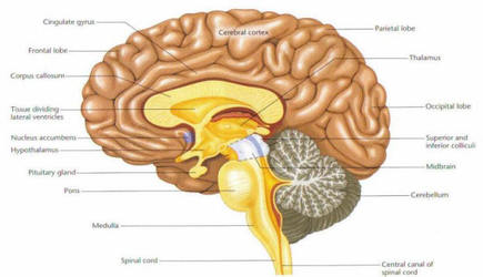
The Human Brain - Visible Body The brain gives us self-awareness and the ability to speak and move in the world. Its four major regions make this possible: The cerebrum, with its cerebral cortex, gives us conscious control of our actions. The diencephalon mediates sensations, manages emotions, and commands whole internal systems. The cerebellum adjusts body movements, speech ... Diagram Of Brain with their Labelings and Detailed Explanation The parietal lobe is found at the upper back of our brain. This lobe functions by controlling all our complex behaviours, including senses of vision, the sense of touch, spatial orientation and body awareness. It manages body position, movements, the perception of stimuli, orientation, handwriting and visuospatial processing. The Occipital Lobe
MRI Segmentation of the Human Brain: Challenges, Methods, … 01.03.2015 · If an atlas or template of the human brain for a specific population of interest is available, then atlas-based methods can be a powerful tool for brain MRI segmentation. The atlas contains information about the brain anatomy (e.g., it contains the information about the location of different brain structures) and it is used as a reference (a prior knowledge) for segmenting …

Labels of the human brain
Frontiers | 101 Labeled Brain Images and a Consistent Human Cortical ... Labeling the macroscopic anatomy of the human brain is instrumental in educating biologists and clinicians, visualizing biomedical data, localizing brain data for identification and comparison, and perhaps most importantly, subdividing brain data for analysis. Human Brain Photos and Premium High Res Pictures - Getty Images Browse 27,835 human brain stock photos and images available, or search for human brain anatomy or human brain illustration to find more great stock photos and pictures. Related searches: human brain anatomy. human brain illustration. Human brain - Wikipedia The brainstem includes the midbrain, the pons, and the medulla oblongata. Behind the brainstem is the cerebellum ( Latin: little brain ). [8] The cerebrum, brainstem, cerebellum, and spinal cord are covered by three membranes called meninges. The membranes are the tough dura mater; the middle arachnoid mater and the more delicate inner pia mater.
Labels of the human brain. Amazon.com: XINDAM 3D Human Brain with Labels Anatomical Model ... Product Description Package includes:a 3.2 inch crystal glass ball,a colorful LED base,a USB cable. Size:3.2 Inch Made from glass and the amazing power of a laser. It can be used as a teaching tool to show a human anatomical Brain It can be used as an interesting science gift for your love. Product information Warranty & Support The Human Brain Atlas at Michigan State University The Human Brain Atlas Keith D. Sudheimer, Brian M. Winn, Garrett M. Kerndt, Jay M. Shoaps, Kristina K. Davis, Archibald J. Fobbs Jr., and John I. Johnson Radiology Department, Communications Technology Laboratory, and College of Human Medicine, Michigan State University; National Museum of Health and Medicine, Armed Forces Institute of Pathology PDF Label the Brain Anatomy Diagram - Gulf Coast State College from the base of the brain to the hip area, running through the spine (vertebrae). Temporal Lobe of the Cerebrum - the region at the lower side of each cerebral hemisphere; contains centers of hearing and memory (located at the sides of the head). Brain Stem - includes midbrain, pons and medulla oblongata, it connects to the spinal cord to higher File:Brain human normal inferior view with labels en.svg Original upload log []. File:Brain_human_normal_inferior_view.svg licensed with Cc-by-2.5 . 2009-10-13T16:18:05Z Beao 424x505 (209117 Bytes) Replaced right brain half with a clone of left brain half because they look excly the same in the picture.; 2007-09-23T15:14:17Z Ysangkok 424x505 (417241 Bytes) removing credits; 2007-03-03T17:30:01Z Ysangkok 424x505 (417718 Bytes) trying to make it work ...
Sex beyond the genitalia: The human brain mosaic | PNAS 30.11.2015 · Previous criticisms of the dichotomous view of human brain have focused on the fact that most sex/gender differences are nondimorphic population-level differences with extensive overlap of the distributions of females and males and have therefore claimed that human brains cannot be sorted into two distinct classes: “male brains” and “female brains” (6–8). Labeled Diagrams of the Human Brain You'll Want to Copy Now The central core consists of the thalamus, pons, cerebellum, reticular formation and medulla. These five regions are the central areas that regulate breathing, pulse, arousal, balance, sleep and early stages of processing sensory information. The thalamus interprets the sensory information and helps determine what is good and bad. Anatomical diagrams of the brain - e-Anatomy - IMAIOS 13.09.2021 · These anatomical charts include the main diagrams necessary for medical students, nursing students, residents, practitioners, anatomists to study the anatomy of the brain, to illustrate a course or explain a pathology to a patient. This atlas of human anatomy is composed of several chapters: Labeled brain anatomy Images, Stock Photos & Vectors - Shutterstock Find Labeled brain anatomy stock images in HD and millions of other royalty-free stock photos, illustrations and vectors in the Shutterstock collection. Thousands of new, high-quality pictures added every day.
Parts of the Human Brain | Anatomy & Function - Study.com The parts of the brain include the cerebrum, the cerebellum, the brain stem, and the pituitary gland. The brain structure is protected by the skull, which is composed of the cranium and the bones... Human Brain Anatomy - Components of Human Brain with Images Composed of the right and left hemispheres, it is the largest part of the brain and is responsible for the processing of speech, learning, reasoning, emotions, muscular contractions as well as the interpretation of sensory data related to hearing, vision and touch. ii. Cerebellum—the Sub-Cerebral Region: PDF The Human Brain Diagram - Therapist Aid The Human Brain Author: Therapist Aid LLC Created Date: 8/3/2020 5:10:53 PM ... Brain (Human Anatomy): Picture, Function, Parts, Conditions, and More • The cortex is the outermost layer of brain cells. Thinking and voluntary movements begin in the cortex. • The brain stem is between the spinal cord and the rest of the brain. Basic functions like...

TDP-43 mutant transgenic mice develop features of ALS and frontotemporal lobar degeneration | PNAS
The Human Brainnetome Atlas: A New Brain Atlas Based on … 25.07.2016 · While human brain atlasing is thus not only an endeavor that has been ongoing for more than a century but also one that will see constant changes and refinement, the current Brainnetome Atlas represents an important step in this development by providing the first whole-brain parcellation based on structural (connectivity) information on the basis of a robust cross …
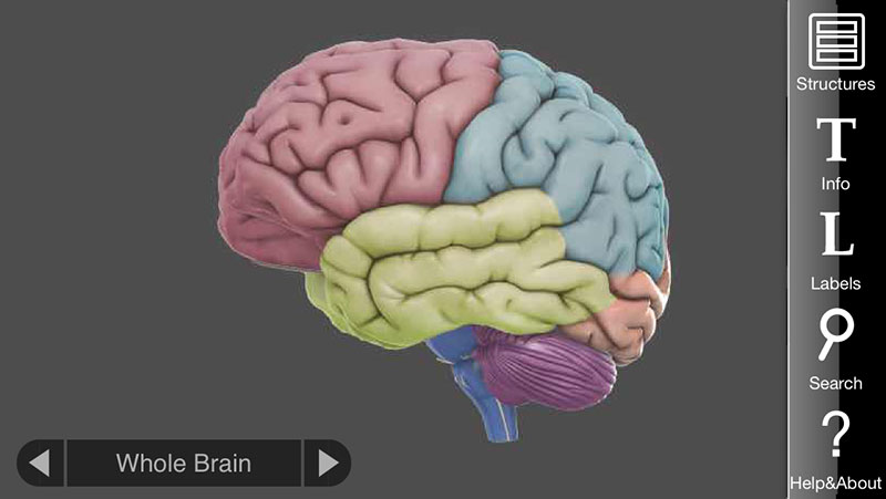
3D Brain App, an interactive way to learn about the different parts of the human brain | Apps ...
101 Labeled Brain Images and a Consistent Human Cortical Labeling ... Labeling the macroscopic anatomy of the human brain is instrumental in educating biologists and clinicians, visualizing biomedical data, localizing brain data for identification and comparison, and perhaps most importantly, subdividing brain data for analysis.
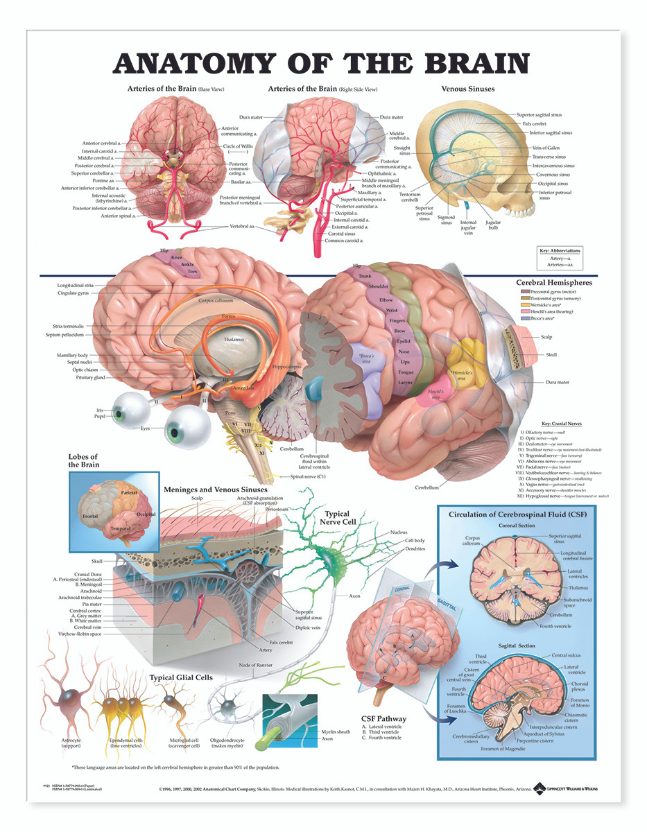
Reference Chart - Anatomy of the Brain - Biologyproducts.com
Labeled Brain Model Diagram | Science Trends The frontal lobe of the brain is responsible for our critical thinking, planning, reasoning, and problem-solving, as well as our experience of emotions. The rear portion of the frontal lobe is the motor cortex, which receives inputs from the other lobes and carries out the movements of the body associated with them.
Brain Viewed from Above | ClipArt ETC
human anatomy labeled human anatomy labeled. Cranial nerves brainstem human inferior cadaver brain swallowing anatomy nerve anterior study crazy. Eye anatomy notes 21 ideas in 2020. Muscle thigh anatomy human muscles leg labeled lower body posterior hip legs physiology origins systems. Toad Dissection (how to/how not to) - YouTube we have 9 Images about Toad ...

Human Anatomy Lab: Muscles of the Arm
Nervous System - Label the Brain - TheInspiredInstructor.com Nervous System - Label the Brain Nervous System - Brain Name: Choose the correct names for the parts of the brain. ( 1) (2) (3) (4) (5) (6) (7) (8) ( 9) This brain part controls thinking. (10) This brain part controls balance, movement, and coordination. (11) This brain part controls involuntary actions such as breathing, heartbeats, and digestion.
Creative Brain Drawing | Free download on ClipArtMag
Brain Anatomy and How the Brain Works - Hopkins Medicine Gray and white matter are two different regions of the central nervous system. In the brain, gray matter refers to the darker, outer portion, while white matter describes the lighter, inner section underneath. In the spinal cord, this order is reversed: The white matter is on the outside, and the gray matter sits within.
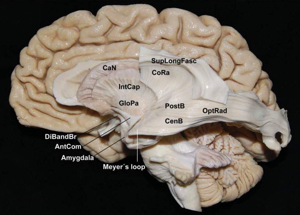
Lateral View of Optic Radiation of Left Hemisphere | Neuroanatomy | The Neurosurgical Atlas, by ...
Label the structures of the human brain midsagittal | Chegg.com Expert Answer. Transcribed image text: Label the structures of the human brain midsagittal (median) section by clicking and dragging the labels to the correct location. Medulla oblongata Infundibulum Pons Hypothalamus Thalamus Fornix Corpus callosum Pituitary gland Midbrain Arbor vitae Mammillary body Septum pellucidum Pineal gland m have & Ext.
Drugs, NTs and the Brain
Brain Basics: Know Your Brain | National Institute of Neurological ... The brain can be divided into three basic units: the forebrain, the midbrain, and the hindbrain. The hindbrain includes the upper part of the spinal cord, the brain stem, and a wrinkled ball of tissue called the cerebellum ( 1 ). The hindbrain controls the body's vital functions such as respiration and heart rate.

12.5 Label The Brain
The Brain and Nervous System | Noba There are approximately 86 billion neurons in the human brain and each has many contacts with other neurons, ... [Image: Biology Corner, , CC-BY-NC-SA 2.0, , labels added] The cerebrum (also called the “cerebral cortex”) is the “newest,” most advanced portion of the brain. The cerebral hemispheres (the left and right …
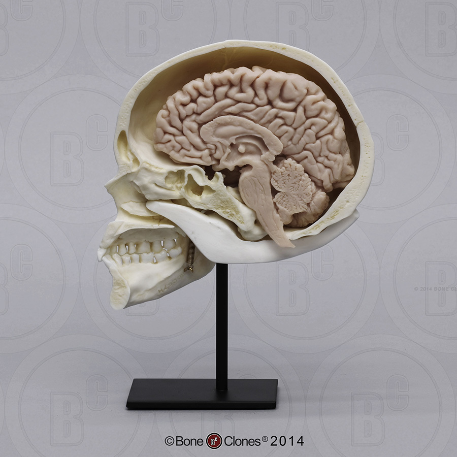
Human Sagittal Cut Half Skull with Brain Hemisphere - Bone Clones, Inc. - Osteological Reproductions
Main Parts of the Human Brain and Subdivisions of Human Brain Parts Human encephalon resembles, in structure and function, with that of the other vertebrates. The scientists reveal that parts of the human brain are Forebrain, Midbrain & Hindbrain and the related structures that collectively act as a single highly specialized unit. These parts work in coordination and perform different functions of brain. More ...

Anatomy of the Brain Neurology Education Poster 24x36 – BananaRoad
Amazon.com: brain model labeled VEVOR Human Brain Model Anatomy 4-Part Model of Brain w/Labels & Display Base Color-Coded Life Size Human Brain Anatomical Model Brain Teaching Human Brain for Science Classroom Study Display Model 3 $159 19 Get it Wed, Mar 30 - Mon, Apr 4 FREE Shipping

Question 3 | Revision World
Labeled Parts Of The Brain Illustrations, Royalty-Free Vector ... - iStock detailed anatomy of the human brain. Illustration showing the medulla, pons, cerebellum, hypothalamus, thalamus, midbrain. Sagittal view of the brain. Isolated on a white background. Diagram of a Brain Colored and labeled human brain diagram Colored and labeled human brain diagram. Flat vector illustration.
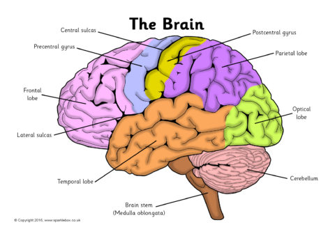
Label the Brain Worksheets (SB11585) - SparkleBox
The Tale of the Dueling Neurosurgeons: The History of the Human Brain ... Early studies of the human brain used a simple method: wait for misfortune to strike -- strokes, seizures, infectious diseases, horrendous accidents -- and see how victims coped. In many cases their survival was miraculous, if puzzling. Observers were amazed by the transformations that took place when different parts of the brain were destroyed, altering victims' personalities. …
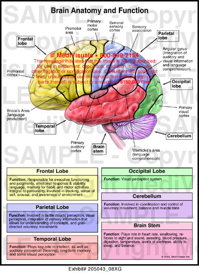
Brain Anatomy and Function Medical Illustration Medivisuals
Brain charts for the human lifespan | Nature 06.04.2022 · We created brain charts for the human lifespan using generalized additive models for location, scale and shape 2,24 (GAMLSS), a robust …
Radiology - Normal brain anatomy - CT and MRI - YouTube
Label the Human Brain - 4th Grade Science Worksheet - SoD Your brain helps your body to run smoothly and controls everything you do, asleep or wide awake. This science worksheet for 4th grade helps you learn about its different parts. Label the Human Brain - 4th Grade Science Worksheet - SoD

68 best images about neuro on Pinterest | Charts, Neuroplasticity and Brain structure
Brain (CNS) Cell Types: Neurons, Astrocytes, Microglia, Human brain tissue was obtained postmortem from patients who had been treated with the thymidine analog, bromodeoxyuridine (BrdU), that labels DNA during the S phase. Using immunofluorescent labeling for BrdU and for one of the neuronal markers, NeuN, calbindin or neuron specific enolase (NSE), we demonstrate that new neurons, as defined by these …
Post a Comment for "42 labels of the human brain"